Playback starts YouTube’s embedded player (privacy-enhanced mode).
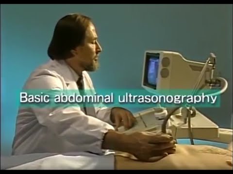
How to do abdominal ultrasound examination
ChannelSonoknowChapters
Description from the creator
Old but still valid information.
0:00 - Intro
1:07 - Ultrasound fundamentals
3:12 - Preparations for examination
6:35 - Scanning basics
8:48 - How images are displayed on monitor
10:55 - 1 Basic abdominal scan
11:21 - 2 Left intercostal scan (spleen, left kidney)
14:58 - 3 Longitudinal scan (left hepatic lobe, pancreas)
18:40 - 4 Transverse scan (pancreas)
21:20 - 5 Oblique scan (tail of pancreas)
22:26 - 6 Right subcostal margin scan (liver, pv, rhv, ivc, right kidney, lhd, gall bladder)
30:02 - 7 Right hypochondriac oblique scan (trunk of pv, sbd, ibd)
31:26 - 8 Right intercostal scan (right hepatic lobe, gall bladder, rhd, right kidney, Morrison's pouch)
33:29 - Image adjustment and recording
rhv = right hepatic vein
rhd - right hepatic duct
pv = portal vein
ivc = inferior vena cava
lhd = left hepatic duct
sbd = superior bile duct
ibd = inferior bile duct
0:00 - Intro
1:07 - Ultrasound fundamentals
3:12 - Preparations for examination
6:35 - Scanning basics
8:48 - How images are displayed on monitor
10:55 - 1 Basic abdominal scan
11:21 - 2 Left intercostal scan (spleen, left kidney)
14:58 - 3 Longitudinal scan (left hepatic lobe, pancreas)
18:40 - 4 Transverse scan (pancreas)
21:20 - 5 Oblique scan (tail of pancreas)
22:26 - 6 Right subcostal margin scan (liver, pv, rhv, ivc, right kidney, lhd, gall bladder)
30:02 - 7 Right hypochondriac oblique scan (trunk of pv, sbd, ibd)
31:26 - 8 Right intercostal scan (right hepatic lobe, gall bladder, rhd, right kidney, Morrison's pouch)
33:29 - Image adjustment and recording
rhv = right hepatic vein
rhd - right hepatic duct
pv = portal vein
ivc = inferior vena cava
lhd = left hepatic duct
sbd = superior bile duct
ibd = inferior bile duct
About this tutorial
Sonoknow published this 40:35 video tutorial on October 29, 2016. It has been watched 470,457 times on YouTube.
- Channel
- Sonoknow
- Category
- Science & Technology
- Duration
- 40:35
- Published
- October 29, 2016
- Views
- 470,457
- Likes
- 7,142
Share
This tutorial in other languages
- FRComment faire une échographie abdominale
- ESCómo hacer una ecografía abdominal
- PTComo fazer exame de ultra-som abdominal
- ITCome fare esame ecografico addominale
- DEWie Bauchultraschalluntersuchung zu tun
- NLHoe kan abdominale echo-onderzoek doen
- RUКак сделать брюшной УЗИ
- PLJak zrobić brzusznej USG
- JA腹部超音波検査を行う方法
- ARكيفية القيام الفحص بالموجات فوق الصوتية في البطن
- HIपेट अल्ट्रासाउंड परीक्षा कैसे करना है
- IDBagaimana melakukan pemeriksaan USG abdomen